Intelligent Dental Cavity Detection & Classification System
تفاصيل العمل
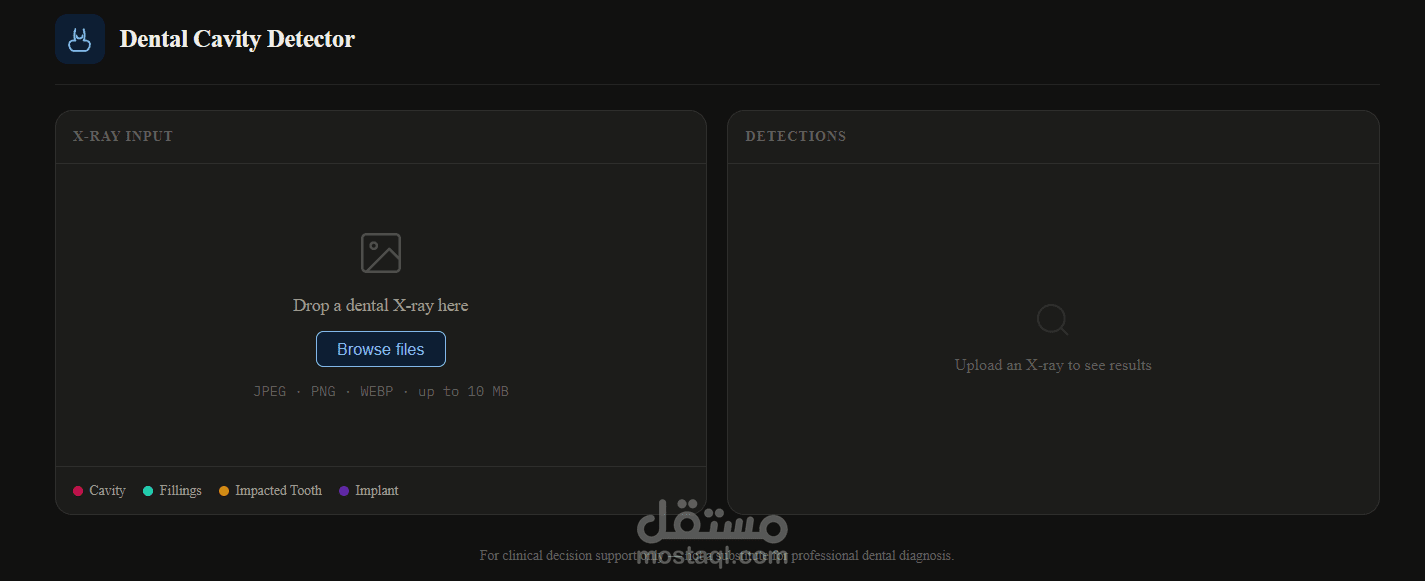
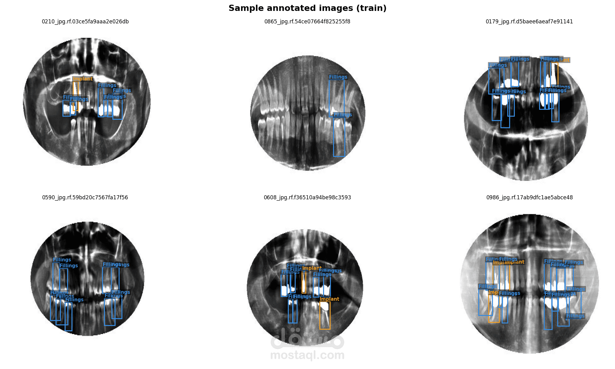
Project Overview:
Developed an advanced, end-to-end Computer Vision pipeline designed to automate the detection and classification of dental cavities from panoramic and bitewing X-ray images. This project demonstrates deep expertise in medical image analysis, combining state-of-the-art object detection with custom-built classification networks to deliver highly accurate diagnostic insights.
My Role & Core AI Contributions:
I led the entire machine learning lifecycle, focusing heavily on architectural design, precise medical data handling, and rigorous performance evaluation.
Medical Image Preprocessing Pipeline: Engineered robust preprocessing techniques specifically tailored for dental radiography. This included contrast enhancement, noise reduction, and data augmentation to handle the complexities and variations inherent in medical X-rays, ensuring the models received high-signal input data.
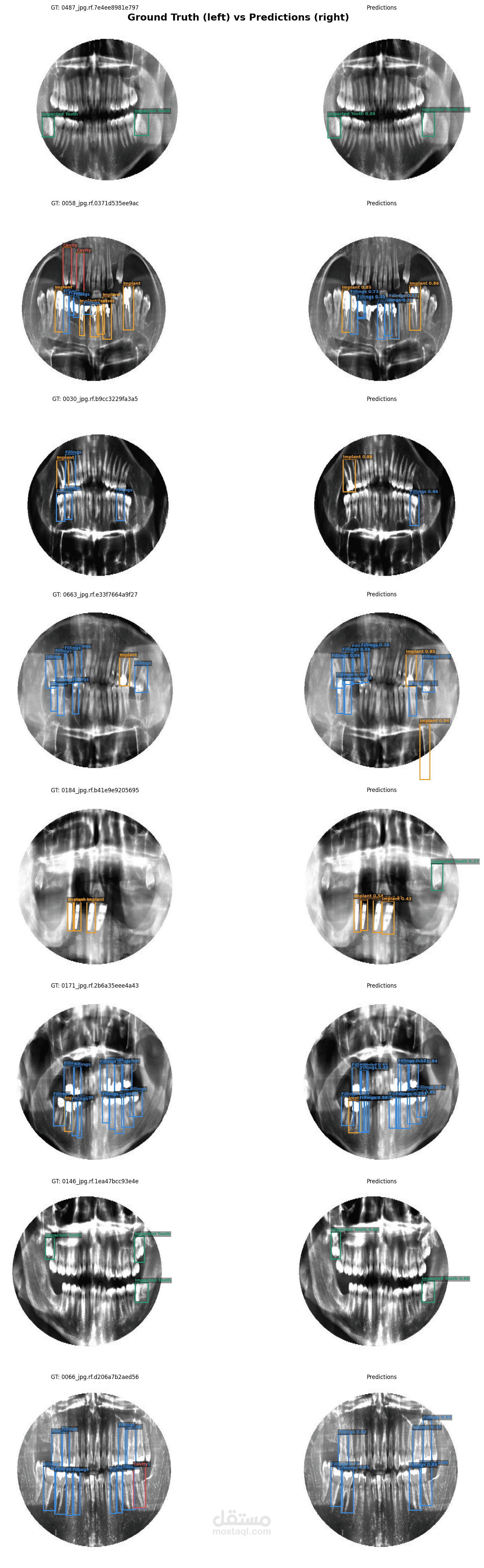
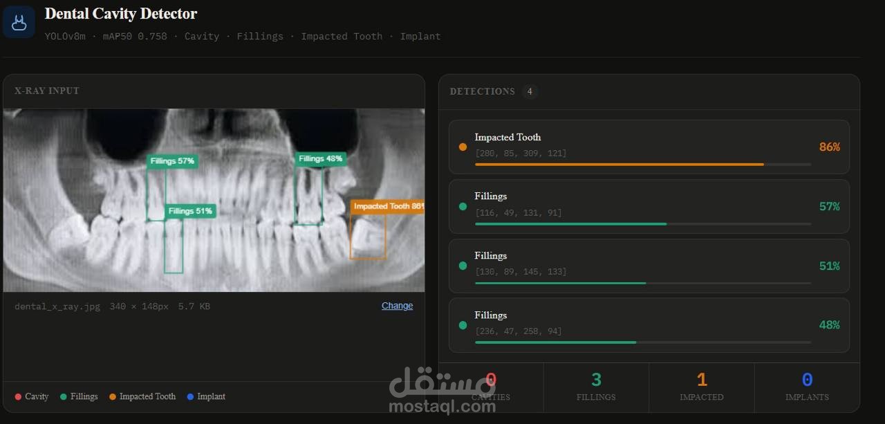
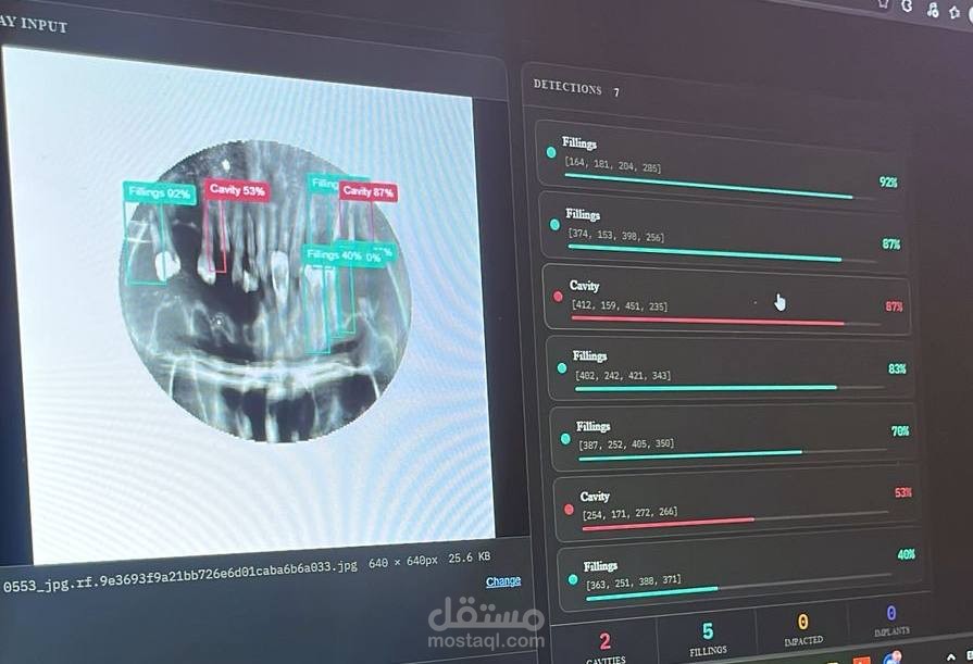
High-Precision Object Detection (YOLOv8): Implemented, configured, and fine-tuned the YOLOv8 architecture to accurately localize cavities. The model efficiently scans complex dental structures and outputs precise bounding boxes around infected areas.
Custom CNN Architecture Design: Engineered and trained custom Convolutional Neural Network (CNN) architectures from scratch to classify the detected regions. This required deep architectural optimization to balance computational efficiency with high classification accuracy.
Comprehensive Model Evaluation: Conducted rigorous technical analysis and benchmarking of the model's performance. Generated detailed analytics, including Accuracy/Loss graphs and Confusion Matrices, to validate the system's reliability and scientific rigor.
Tech Stack & Tools:
Python, Computer Vision, Deep Learning, YOLOv8, Custom CNNs, Medical Image Processing, Data Augmentation, Performance Analytics.