brain tumor detection
تفاصيل العمل
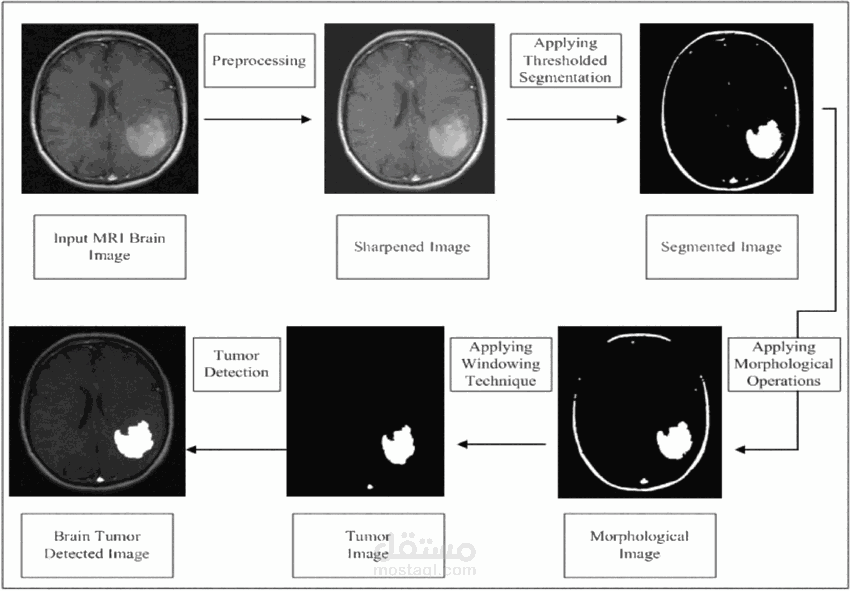


Detecting brain tumors using Convolutional Neural Networks (CNNs) is a fascinating application of deep learning in medical imaging. Here's an overview of how it works:
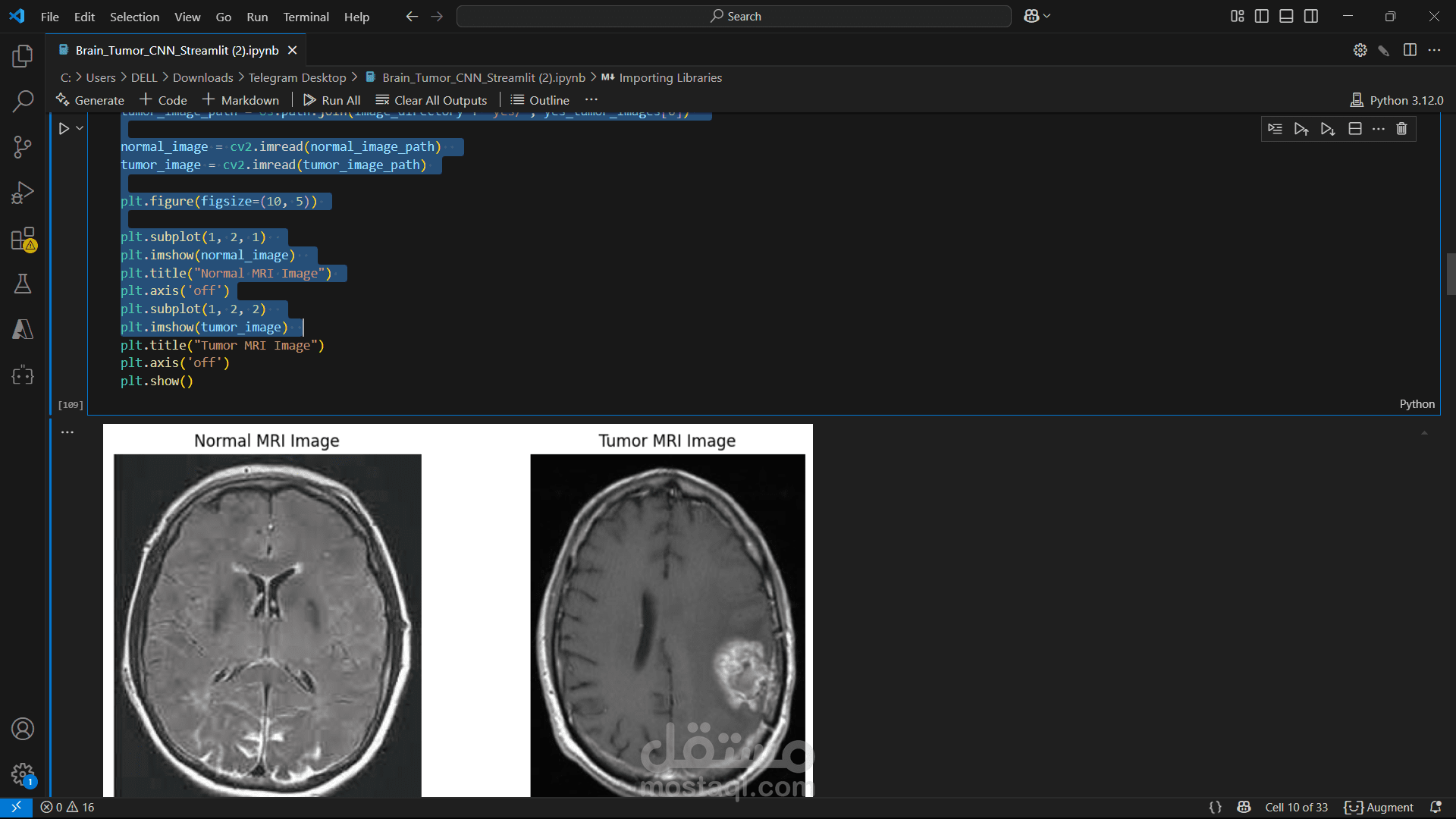
Data Collection: The process begins with collecting a large dataset of brain MRI images. These images are labeled to indicate whether they contain a tumor or not, and sometimes even the type of tumor.
Preprocessing: The MRI images are preprocessed to enhance their quality and make them suitable for training the CNN. This includes steps like resizing the images, normalizing pixel values, and augmenting the data to increase the diversity of the training set.
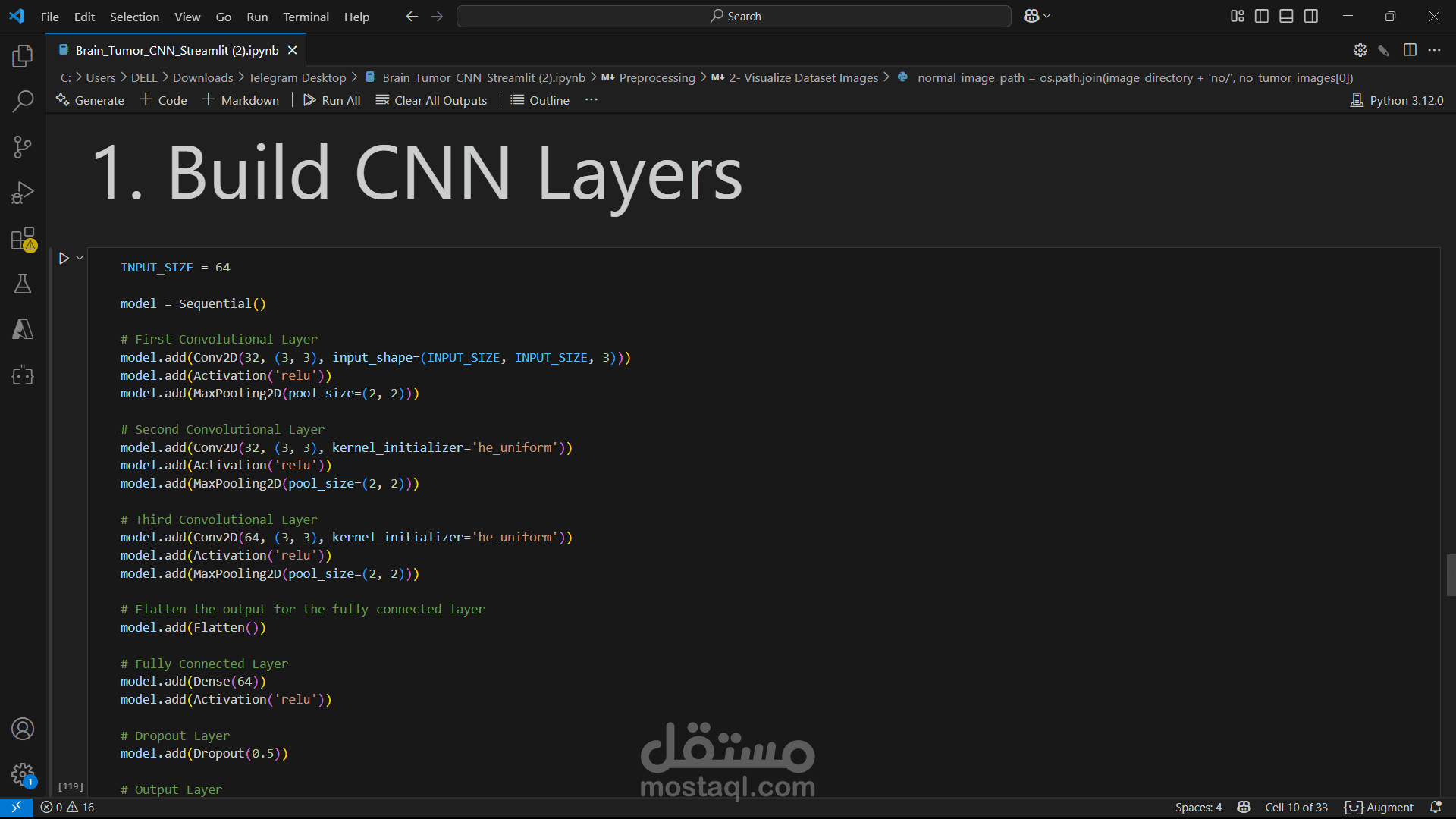
CNN Architecture: A CNN model is designed with multiple layers, including convolutional layers, pooling layers, and fully connected layers. The convolutional layers extract features from the images, such as edges and textures, while the pooling layers reduce the spatial dimensions, and the fully connected layers perform the final classification.
Training: The CNN model is trained using the labeled MRI images. During training, the model learns to recognize patterns and features associated with brain tumors. The training process involves optimizing the model's parameters to minimize the difference between the predicted and actual labels.
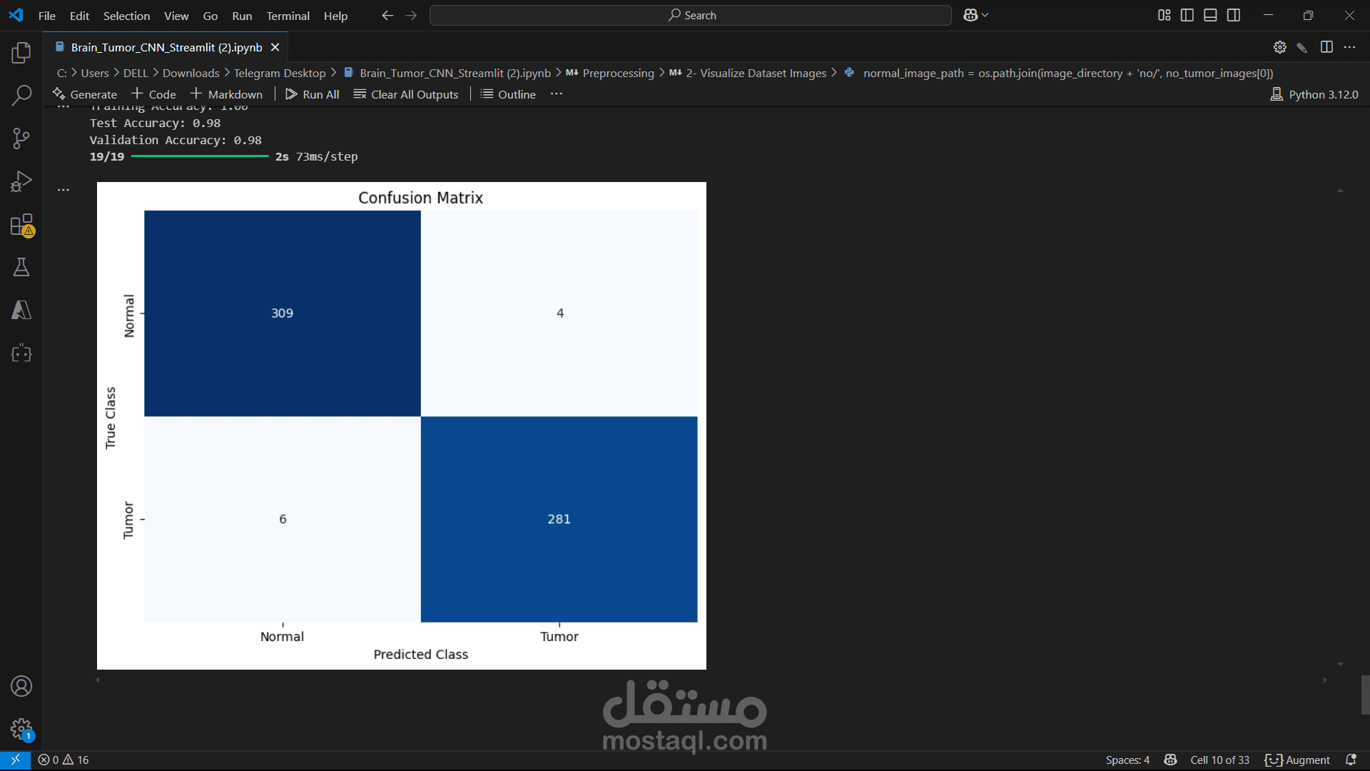
Evaluation: The trained model is evaluated using a separate set of MRI images that were not used during training. Metrics like accuracy, precision, recall, and F1-score are used to assess the model's performance.
Prediction: Once the model is trained and evaluated, it can be used to predict the presence of brain tumors in new MRI images. The model outputs a probability score indicating the likelihood of a tumor being present.
Deployment: The trained CNN model can be deployed in a clinical setting to assist radiologists in diagnosing brain tumors. It can provide quick and reliable predictions, helping to improve the accuracy and efficiency of medical diagnoses.